Brainstem injuries


The brain stem (truncus cerebri) is located deep in the brain and provides the connection with the spinal cord. The brain stem serves as a connection between the brain and the body, coordinating motor control signals sent from the cerebrum to the spinal cord. It consists of the medulla oblongata, the pons and the midbrain, (mesencephalon).
The hypothalamus and pituitary gland also belong to the brainstem.
The brainstem monitors and regulates the autonomic nervous system; breathing, heart rate, blood pressure and circulation, chewing and swallowing, reflexes of seeing and hearing (startle) sweating, digestion, body temperature, pupil size.
It affects alertness, tasting, formation of saliva, vomiting, urination, ability to sleep (sleep-wake cycle regulation), sense of balance (vestibular), feeling of gravity and can cause a central sleep apnea (CSAS),
Scattered throughout the brainstem, is the reticular formation that controls consciousness (that distinguishes important and unimportant stimuli)
Observed problems:
Diminished vital capacity in the respiration, which is important for speech
Difficulty in swallowing food and water (Dysphagia)
Problems with balance and movement
Dizziness and nausea (Vertigo)
Sleep problems (insomnia, sleep apnea).
All of the vital functions mentioned above can be damaged and can give a direct life threatening danger.
The brain stem plays a vital role in attention, arousal, and consciousness. All information to and from the body passes through the brain stem on its way to or from the brain.
Like the frontal and temporal lobes, the brain stem is located in an area near bony protrusions making it vulnerable to damage during an accident.
When a person's brain stops functioning, this state is called brain death. In other states with loss of consciousness such as coma, the brain still works.
Parts of the brainstem
Medulla Oblongata
Medulla oblongata is located at the bottom of the brain stem. It is the part of the brain that you absolutely don't want to injure, since it plays a major role in the vital functions of the brain stem. Medulla oblongata regulates vital characteristics of the body, such as blood pressure, heartbeat, breathing, sleep cycles, and digestion. It is also responsible for reflexes, especially reflexes of the face and throat (blinking, coughing, sneezing, and gagging), motor control, and certain senses, such as touch.
Pons
Pons is a bulge, situated between the medulla and the midbrain, right in front of the cerebellum. It consists of large bundles of nerve fiber, which connect each side of the cerebellum to the opposite cerebral hemisphere. Pons mostly serves as a node that relays neural impulses, governing voluntary movement, and transfers information between the medulla oblongata and the cerebral cortex. Pons also relays sensory information and signals governing sleep patterns. If pons is damaged, it may cause loss of all muscle function except for eye movement.
Midbrain
Midbrain is located above the pons, in the upper part of the brainstem. Midbrain consists of the cerebral peduncle, red nucleus, and substantia nigra. Of the functions of brain stem, the midbrain is responsible for voluntary movement, visual/auditory reflexes, and consciousness. The bottom of the midbrain also includes centers responsible for relaying information about pain, temperature, and touch.
Thalamus and Hypothalamus
Thalamus and hypothalamus are situated between the brain stem and the cerebellum, in which the hypothalamus is located directly below the thalamus. The primary function of thalamus, as a part of the brain stem, is to receive all sensory signals except for smell, and to send out motor signals. Hypothalamus, on the other hand, is involved in controlling processes and related drives. The processes include eating, drinking, sleep, and sexual activity. Hypothalamus is also responsible for controlling body temperature and internal organs, and coordinates brain stem activity.
Despite the sections of the brain stem serving different roles, they are connected, both anatomically and functionally. Poms, midbrain, thalamus, hypothalamus, and the midbrain all collaborate so the human body doesn't suffocate while sleeping. Nevertheless, the functions of the brain stem are wider than that: brain stem controls involuntary functions such as breathing, heartbeat, sleeping, digestion, appetite, sexual drive, voluntary movements, and reflexes. It transmits signals from body to the brain, and even plays a role in higher cognitive functioning.
A brainstem infarction is called, depending on the area affected, syndrome of:
- Avellis (in the medulla oblongata)
- Benedict (in the midbrain/mesencephalon).
- Claude (in the midbrain/mesencephalon)
- Foville (in the pons, on the back (dorsal) of the pontine tegmentum)
- Millard-Gubler (in the pons, also called ventral pontine syndrome)
- Wallenberg (in the medulla oblongata)
- Weber (in the midbrain/mesencephalon)
Benedikt's syndrome (Benedict)
As seen with Weber syndrome, people with this syndrome have a palsy or a waddling walk through coordination disorder (ataxia). Slight paralysis or weakness occurring on the side of the body opposite tot the side of the brain in which the causal lesion occurs. But also involuntary vibrations which occur when performing a deliberate operation (intention tremor), and abnormal involuntary movements (hyperkinesias).
Claude's syndrome
Claude's syndrome is very rare. The syndrome was first described by the French neurologist and psychiatrist H.C.J. Claude.
Symptoms include:
- loss/paralysis on the side of the lesion (ipsilateral) of the oculomotor nerve. This is the third cranial nerve; the oculomotor nerve (nIII)
- a half-sided loss (hemiparesis) of the shoulder, tongue and lower part of the face on the other side of the body (contralateral)
- a coordination disorder and difficulty with articulation (dysarthria).
The coordination disorder is caused by damage to the red nucleus, by the fact that it cannot receive input from the cerebellum.
Foville syndrome
Foville syndrome is caused by damage to the side branches of the basilar artery in the area of the pons. The pons is the middle part of the brain stem. It can be caused by a tumor, by a CVA / stroke, by traumatic brain injury or by inflammation of the brain. The inflammation can also be caused by abscesses in the body, for example a lung abscess.
Other names are:
Foville paralysis, Foville bridge syndrome, Caudal bridge hood syndrome, hemiplegia abducento-facialis alternans, hemiplegia alternansinferieure pontina.
Symptoms:
- On the side of the injury, there is loss or paralysis of the eye muscle, causing the eye to be directed to one side (ipsilateral horizontal gaze paresis or gaze paralysis). Gaze paralysis means that someone cannot move their eyes in the same direction at the same time. With horizontal gaze palsy, a person cannot look to the left or right. Weakness of horizontal eye movements (internuclear ophthalmoplegia)
- Facial paralysis on one side, on the side of the brainstem lesion (ipsilateral facial nerve palsy)
- On the other side of the lesion, there is loss of sensation or paralysis in the limbs (contralateral hemiplegia/hemiparesis)
- On the other side of the lesion, there is loss of sensation on one side of the face (contralateral hemisensory loss). Complete or partial loss of sensation of heat perception.
For professionals: Foville syndrome manifests itself with ipsilateral sixth nerve palsy, facial paralysis, and contralateral hemiparesis.
There are reports of other features such as facial hypoesthesia, peripheral deafness, Horner syndrome, ataxia, pain, and thermal hypoesthesia.
Millard Gubler Syndrome
The Millard Gubler syndrome is also called ventral pontine syndrome.
Symptoms:
- On the side of the injury, there is loss or paralysis (ipsilateral paresis) of one of the six eye muscles that can move the eye.
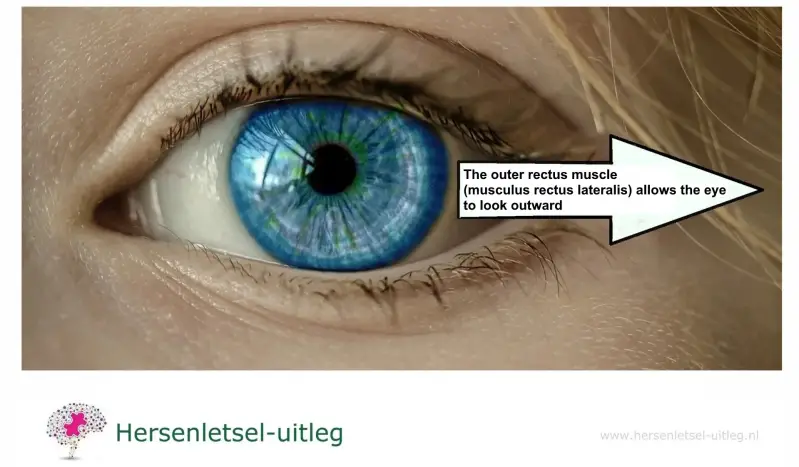
This eye muscle, on the side of the eye towards the cheek (musculus rectus lateralis), normally ensures that someone can move the eye outwards, towards the ear (abduction).
The paralysis of this eye muscle is caused by damage to cranial nerve VI, resulting in the muscle no longer being able to turn the eye outwards and the person seeing everything double (diplopia).
- On the side of the injury, there is loss or paralysis (ipsilateral paresis) of the upper and lower face (damage to cranial nerve VII).
- On the other side of the injury, there is one-sided paralysis (contralateral hemiplegia) of the upper and lower limbs. This is due to damage to the pyramidal tract (corticospinal tract). Millard-Gubler syndrome can lead to spastic disorders, which means that the limbs can only be moved to a very limited extent.
Each eye has six muscles. Cooperation between the muscles of the eyes allows the eyes to see in a coordinated manner and in this way double vision is prevented.

Brain damage can cause an eye muscle to fail. Then the muscle cannot cooperate with other muscles. As a result, the images of the two eyes do not coincide.
The causes of this syndrome often vary with age. In young people, the syndrome occurs due to tumors, infectious diseases (neurocysticercosis and tuberculosis), demyelinating diseases (multiple sclerosis) or a viral infection that causes inflammation of the brain (Rhomb encephalitis). In older people, a cerebral infarction or cerebral hemorrhage can be the cause. There are also known patients with a rupture of the basilar artery, an aneurysm of the arteria basilaris.
Wallenberg syndrome (Lateral medullary syndrome)
Cerebral infarction or hemorrhage (stroke) in the medulla in the brainstem, has been named specifically as the syndrome of Wallenberg (or Wallenberg syndrome).
The characteristic features are: suffer from hiccups, acute vertigo, jerky eye movements (nystagmus), difficulty swallowing, vomiting, and disorder in the vocalisation, including dysarthria and hoarseness.
It depends on the location if half-sided sweat secretion is reduced (sometimes even more!) Sensory disturbance in the soft palate. The Horner's syndrome is seen in this syndrome: a drooping eyelid and a pupil is smaller. People with this syndrome may be on one side of the face feel less (pain and temperature) and on the other side of their body too. Tinnitus (ringing) is a specified complaint.
Most people with the Wallenberg syndrome recover better than people who have had a different type of stroke. The recovery differs from one person to another: from six weeks to six months and others with more significant damage may have a more permanent disability.
 Horner's syndrome
 Nystagmus
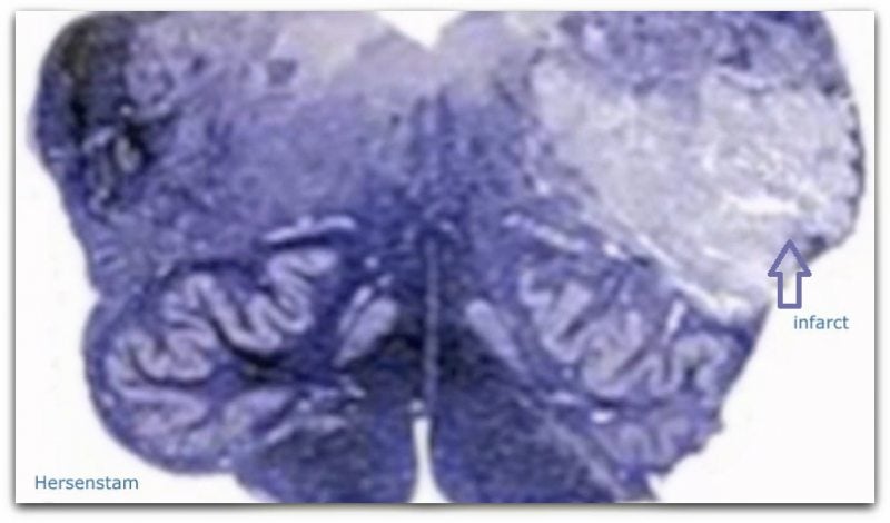
 Brainstem infarctation (see arrow)
In summary:
- hoarseness (dysphonia)
- difficulty to articulate (dysarthria)
- nausea and vomiting
- hiccups
- rapid eye movements (nystagmus)
- a decrease in sweating (sometimes increase!)
- half sided feeling disorder -pains and temperature (sensory disturbance) perceived difference in how hot or cold something is on one side of the body
- dizziness
- difficulty in walking
- problems with the balance to be maintained
- sometimes sided paralysis or loss of strength and sensation (in arm, trunk, leg, face or tongue)
- loss of taste in the half of the tongue
- complaints that everything around the patient seems tilted or unbalanced
- heart rate deceleration (bradycardia)
- high or low blood pressure
- a co-ordination disorder (ataxia) at one side of the body (hemiataxia)
- vocal cord paralysis
- photosensitivity
- neuropathic pain (nerve pain)
Weber's syndrome
This syndrome is characterized by spastic half sided slight paralysis or weakness in the body (hemiparesis), loss of facial muscles with the exception of the upper part of the facial muscles and a half sided failure (hemipareses)of the tongue.
The eye symptoms in this syndrome are a wide, light stiff pupil and the eye can not move from one side tot the other.
Resources
Team Hersenletsel-uitleg
Brogna C, Fiengo L, Türe U. Achille Louis Foville's atlas of brain anatomy and the Defoville syndrome. Neurosurgery. 2012 May;70(5):1265-73; discussion 1273. [PubMed] [Reference list]
Canepa Raggio C, Dasgupta A. Three cases of Spontaneous Vertebral Artery Dissection (SVAD), resulting in two cases of Wallenberg syndrome and one case of Foville syndrome in young, healthy men. BMJ Case Rep. 2014 Apr 28;2014 [PMC free article] [PubMed] [Reference list]
Claude H, Loyez M (1912). "Ramollissement du noyau rouge". Rev Neurol (Paris). 24: 49–51. Haines, Duane.E. Neuroanatomy: An Atlas of Structures, Sections, and Systems, Volume 153,Nummer 2004 Frumkin LR, Baloh RW. Wallenberg's syndrome following neck manipulation. Neurology. 1990 Apr;40(4):611-5. doi: 10.1212/wnl.40.4.611. PMID: 2181339
Massi DG, Nyassinde J, Ndiaye MM. Superior Foville syndrome due to pontine hemorrhage: a case report. Pan Afr Med J. 2016;25:215. [PMC free article] [PubMed] [Reference list]
Menéndez-González M, García C, Suárez E, Fernández-Díaz D, Blázquez-Menes B. Sindrome de Wallemberg secundario a disección de la arteria vertebral por manipulación quiropráctica [Wallenberg's syndrome secondary to dissection of the vertebral artery caused by chiropractic manipulation]. Rev Neurol. 2003 Nov 1-15;37(9):837-9. Spanish. PMID: 14606051.
Raghachaitanya Sakuru; Ayman G. Elnahry; Pradeep C. Bollu. Millard Gubler Syndrome https://www.ncbi.nlm.nih.gov/books/NBK532907/
Wenban AB. Síndrome de Wallemberg secundario a disección de la arteria vertebral por manipulacion quiropráctica [Wallenberg's syndrome secondary to dissection of the vertebral artery caused by chiropractic manipulation]. Rev Neurol. 2004 Sep 1-15;39(5):497; author reply 497. Spanish. PMID: 15378467. https://books.google.nl/books?id=xP-kmKiziAQC&pg=PA34&hl=nl&source=gbs_selected_pages&cad=2#v=onepage&q&f=false
https://www.ncbi.nlm.nih.gov/books/NBK554418/
ISBN 9031392162 , Neurologie A. Hijdra & P.J. Koudstaal, R.A.C. Roos ISBN 9035234715 , The PICA syndrome, Timothy C. Hain, MD Hossain, M. (2010). Lateral Medullary Syndrome (Wallenberg’s Syndrome) - A Case Report.Faridpur Med. Coll. J., 5(1), 35-36. http://wiki.cns.org/wiki/index.php/Brain_stem_vascular_syndromes
NINDS Wallenberg's Syndrome Information Page. (2007, February 15). National Institute of Neurological Disorders and Stroke. Retrieved June 10, 2013, from http://www.ninds.nih.gov/disorders/wallenbergs/wallenbergs.htm