Gyrus Fusiformis
The function of the gyrus fusiformis
The fusiform gyrus is a part of the brain located at the bottom of the temporal lobe. This area is necessary for recognizing and distinguishing faces, objects, and visual patterns (recognizable images or shapes). It also helps with processing colors and reading words.
In summary:
- Recognizing and reading words
- Facial recognition (prosopagnosia)
- Recognizing emotions in faces
- Body recognition
- Recognizing and distinguishing patterns and objects (visual agnosia)
- Color processing
Consequences of damage to the fusiform gyrus
As may be expected from the above list of functions of the fusiform gyrus, problems occur in specific functions when damaged.
One of the most common consequences is prosopagnosia, also known as face blindness, in which someone has difficulty recognizing faces, even of people they know well. Sometimes a face appears distorted.
Someone with damage to the fusiform gyrus may also no longer be able to recognize facial expressions or emotions. This can cause significant difficulty in interacting with people.
Other possible effects include difficulty identifying objects and patterns (visual agnosia) or difficulty distinguishing small visual details and colors.
Causes of damage to the Fusiform Gyrus
The fusiform gyrus may be damaged by various causes. A common cause is a stroke, where an interruption of the blood supply to this area can lead to loss of function.
Traumatic brain injury, for example from an accident, or surgery on the temporal lobe (temporal lobectomy) may also cause damage. In addition, neurological disorders such as epilepsy, tumors, or degenerative diseases, including Alzheimer's, may negatively impact the fusiform gyrus. Finally, an infection or inflammation in the brain (encephalitis) may also cause damage, potentially impacting cognitive and visual functions.
No damage, but less active fusiform gyrus
Dyslexia
Research shows that in people with dyslexia the fusiform gyrus is less active and contains less gray matter. The severity of word reading difficulties is related to the white matter network, particularly in the right fusiform gyrus.
Autism
Research shows that in people with autism, little to no activity is visible in the fusiform gyrus when viewing a face. This can be explained by the fact that this part of the brain is important for recognizing emotions.
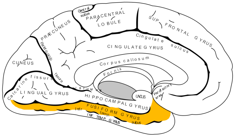
Where in the brain is the fusiform gyrus located?
The fusiform gyrus is an important part of the lateral temporal lobe and the occipital lobe.
The neurologist and anatomist Brodmann numbered the brain areas in 1909 and the fusiform gyrus is called Brodmann area number 37.
It is also called the lateral occipitotemporal gyrus.
The fusiform gyrus is part of the visual processing system.
Together, these areas are responsible for perceiving, interpreting, and understanding visual information such as color, movement, and shape.
Neuropsychological research shows that facial recognition is enabled by an extensive network in the visual part of the brain, or ventral visual pathway. This network extends from the back of the head (lateral occipital sulcus) to the side of the temples (temporal pole).
The fusiform gyrus and the anterior temporal cortex work together to recognize faces. Damage to the anterior temporal cortex makes it difficult to remember faces, while damage to the fusiform gyrus makes it difficult to recognize faces.
In the right hemisphere of the temporal lobe there is an area that allows you to recognize faces as a whole at a glance. The left hemisphere of the brain is responsible for viewing and storing the fine details of faces.
What does face blindness (prosopagnosia) mean in practice?
Persons with face blindness cannot recognize themselves in the mirror. They cannot recognize the faces of partner, children, parents, friends or other acquaintances. They also cannot recognize them in photos. New faces are not remembered. People in uniform or wearing the same outfit all look the same. It makes watching a movie or a play difficult because the characters are unrecognizable.
It is especially difficult if you cannot recognize someone's facial expression. Then you do not really know someone is feeling.
See more on our page about prosopagnosia.
Also read our page about not being able to recognize facial expressions/emotions, and our page about social cognition.
The next page is also important: not being able to recognize objects and patterns. It all has to do with the fusiform gyrus.
Coping with Face Blindness
In rehabilitation clinics, people often learn how to cope with limitations and how to better recognize faces (compensatory strategies). People learn to pay close attention to details such as a mustache, glasses, gait, a specific scent, or hair color. You can also arrange for acquaintances to mention their names when they encounter you or visit. At gatherings with family or friends, you can ask everyone to wear a name tag.
In the Oliver Sacks film, you see how the man who mistook his wife for a hat accidentally reaches for the coat rack, but instead grabs his wife's head. Neurologist Oliver Sacks himself had face blindness.
Watch a video here in which Oliver Sacks explains Face Blindness.
Resources
- Team Hersenletsel-uitleg, team neurologia
- https://www.hersenletsel-uitleg.nl/gevolgen-per-hersengebied/gyrus-fusiformis
- Boller. F & Grafman J. (Eds). (1989). Handbook of Neuropsychology. vol2. Amsterdam Elsevier
- Bruce, V., & Young, A. (1986). Understanding face recognition. British Journal of Psychology, 77, 305–327
- Gallegos, D. R., & Tranel, D. (2005). Positive facial affect facilitates the identification of famous faces. Brain and Language, 93, 338–348.
- Iidaka T. Role of the fusiform gyrus and superior temporal sulcus in face perception and recognition: An empirical review. JapanesePsychological Research. 2014;56 (1): 33-45
- Jonas J, Rossion B, Brissart H, Frismand S, Jacques C, Hossu G, Colnat-Coulbois S, Vespignani H, Vignal JP, Maillard L. Cortex. Beyond the coreface-processing network: Intracerebral stimulation of a face-selective area in the right anterior fusiform gyrus elicits transient prosopagnosia. 2015 Nov;72:140-155. doi: 10.1016/j.cortex.2015.05.026. Epub 2015 Jun 4. PMID: 26143305
- Kanwisher, N. (2000). Domain specificity in face perception. Nature Neuroscience, 3, 759-763.
- Kanwisher N & Yovel G. The Fusiform Face Area: A Cortical Region Specialized for the Perception of Faces. Philos Trans R Soc Lond B Biol Sci.2006;361(1476):2109-28. doi:10.1098/rstb.2006.1934 - Pubmed
- Koh YH. Right Fusiform Gyrus Infarct with Acute Prosopagnosia. Acta Neurol Taiwan. 2022 Dec;31(4):186-187. PMID: 35470413. https://europepmc.org/article/MED/35470413
- Ma Y & Han S. Functional Dissociation of the Left and Right Fusiform Gyrus in Self-Face Recognition. Hum Brain Mapp. 2012;33(10):2255-67.doi:10.1002/hbm.21356 - Pubmed
- Martha J. Farah (1996).Is face recognition 'special'? Evidence from neuropsychology Behavioural Brain Research 76, 181-189
- Mirabella G, Montalti M. When do facial emotions impact inhibitory control and response readiness? A gateway to understanding emotionprocessing. Neurosci Biobehav Rev, 106289, 14 Jul 2025 PMID: 40669527
- Rossion B, Jacques C, Jonas J. The anterior fusiform gyrus: The ghost in the cortical face machine. Neurosci Biobehav Rev, 158:105535, 06Jan 2024
- Rossion B, Caldara R, Seghier M, Schuller A, Lazeyras F, Mayer E. A Network of Occipito-Temporal Face-Sensitive Areas Besides the RightMiddle Fusiform Gyrus is Necessary for Normal Face Processing. Brain. 2003;126(Pt 11):2381-95. doi:10.1093/brain/awg241 - Pubmed
- Srinivasan, R & J. D. Golomb, A. M. Martinez. A Neural Basis of Facial Action Recognition in Humans. Journal of Neuroscience, 2016; 36 (16):4434 DOI: 10.1523/JNEUROSCI.1704-15.2016
- Steeves J, Culham J, Duchaine B et al. The Fusiform Face Area is Not Sufficient for Face Recognition: Evidence from a Patient with DenseProsopagnosia and No Occipital Face Area. Neuropsychologia. 2006;44(4):594-609. doi:10.1016/j.neuropsychologia.2005.06.013 - Pubmed
- Tyler LK, Chiu S, Zhuang J et-al. Objects and categories: feature statistics and object processing in the ventral stream. J Cogn Neurosci.2013;25 (10): 1723-35. doi:10.1162/jocn_a_00419
- https://www.sciencedaily.com/releases/2016/04/160419183920.htm