Blood Vessels
On this page:
Complaints and consequences classified per artery, in case of failure
Names of the arteries in Latin
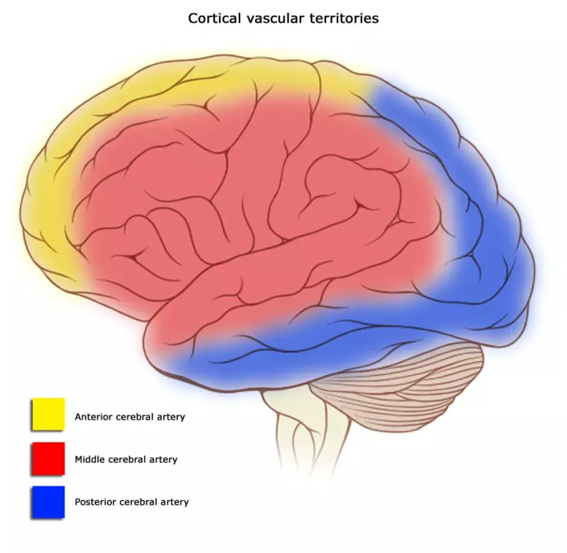
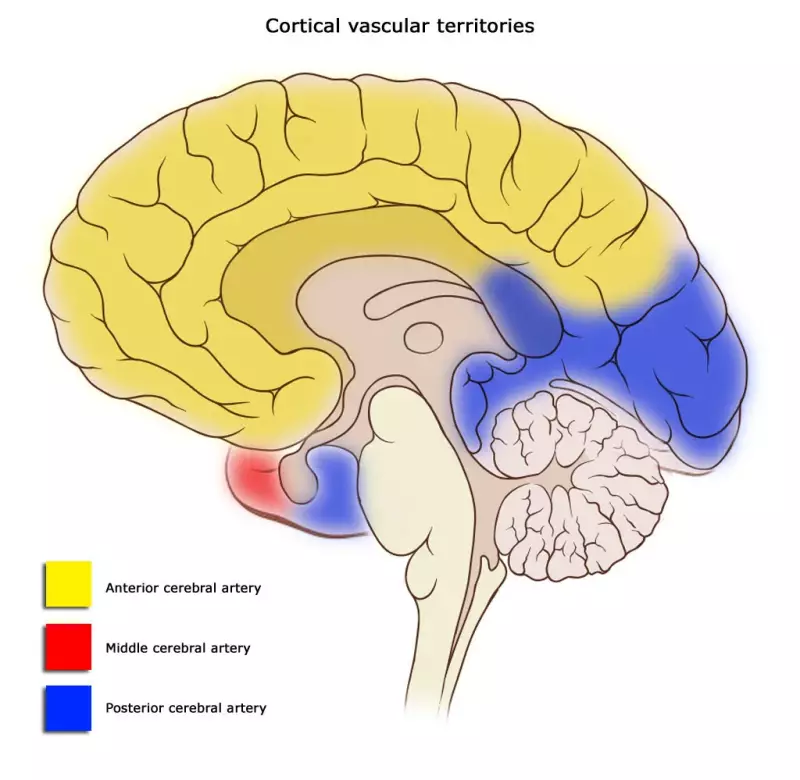
Circulatory area of three cerebral arteries
Two vertebral arteries and their branches (anatomy)
Blood circulation in the skull and scalp (with downloads)
Blood vessels in the brain
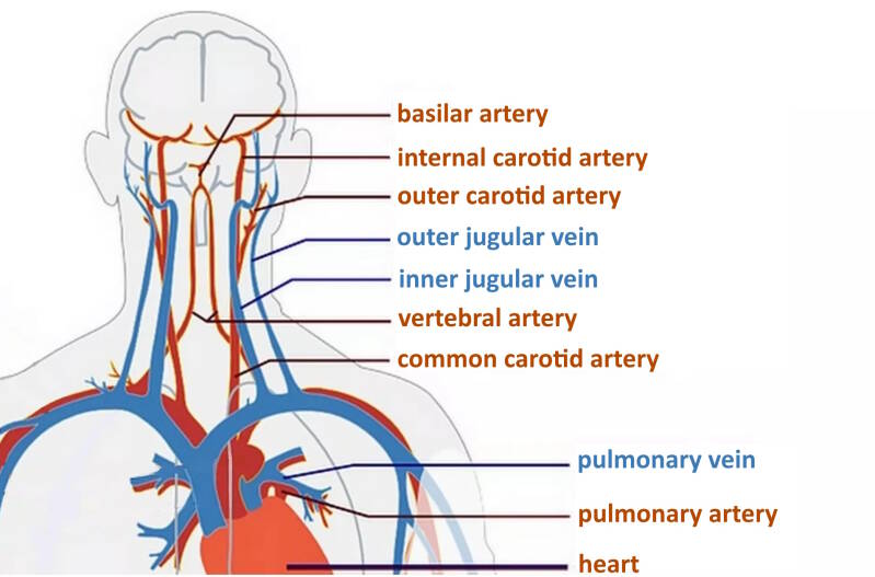
There are two types of blood vessels: the arteries (also called arteries/arteria, abbreviated a.) and the veins (also called veins/vena, abbreviated v.).
A number of large blood vessels, arteries, run from the body artery (aorta) to the brain. Arteries transport oxygen and nutrients to the brain. The brain is supplied with blood through four arteries:
- Two carotid arteries (carotid arteries) at the front of the neck. They supply 80% of the brain with blood and thus with oxygen and glucose.
- Two vertebral arteries / vertebral arteries (arteria vertebralis) that run at the back of the neck along the vertebrae to the brain. They provide the remaining 20% of the blood flow. The vertebral arteries supply blood to the upper part of the spinal cord, the brain stem, the cerebellum and the posterior part of the brain.
The two vertebral arteries together form the basilar artery.
The arteries are colored red in the image below.
Naming
| Name plus abbreviation | Latin |
|---|---|
| Anterior cerebral artery (ACA) | Arteria cerebri anterior |
| Middle cerebral artery (MCA / ACM) | Arteria cerebri media |
| Posterior cerebral artery (ACP) | Arteria cerebri posterior |
| Internal carotid artery (ICA) | Arteria carotis interna |
| Common carotid artery (CCA) | Arteria carotis communis |
| Basilar artery (BA) | Arteria basilaris |
| Vertebral artery (VA) | Arteria vertebralis |
| Posterior cerebral artery (PCA) | Arteria cerebri posterior |
| Posterior communicating artery (PComm) | Arteria communicans posterior |
| Anterior communicating artery (ACOM) | Arteria communicans anterior |
Blood supply to:
| Cerebrum |
Anterior cerebral artery (ACA) Middle cerebral artery (MCA / ACM) Posterior Cerebral artery (ACP) |
| Brainstem |
Posterior cerebral artery (ACP) Basilar artery (BA) Vertebral artery (VA) Superior celebellar artery (SCA) Anterior inferior celebellar artery (AICA) Posterior inferior celebellar artery (PICA) Anterior spinal artery (ASA) |
| Cerebellum / Little brain |
Superior celebellar artery (SCA) Anterior inferior celebellar artery (AICA) Posterior inferior celebellar artery (PICA) |
| Spinal cord |
Anterior spinal artery (ASA) Posterior spinal artery (ASP) |
The two carotid arteries (arteriae carotis) and their branches
The common carotid artery (Arteria carotis communis arises from the aortic arch or a major branch of this arch (truncus brachiocephalis).
This carotid artery branches at the level of the angle of the jaw into:
- Inner carotid artery / Arteria carotis interna,
- Outer carotid artery / Arteria carotis externa

- Arteria carotis externa; This external carotid artery provides blood flow to:
- the thyroid gland
- the larynx
- the tongue
- the face; the facial muscles
- Arteria carotis interna; This internal carotid artery only branches at the base of the skull and makes an S-bend there, then gives a branch to the eye and then branches further into:
- Arteria cerebri anterior (ACA) supplies both large cerebral hemispheres on the medial and dorsal sides of the body with blood from the
The ACA supplies the entire medial part of the brain, i.e. the part for the inside of the body, with blood. the ACA is the least commonly affected by stroke and therefore injury to the ACA can easily be misdiagnosed.
- Middle cerebral artery (ACM) supplies many areas of the brain with blood:
- Frontal lobe
- Temporal lobe
- Parietal lobe
- the ACM branches into the lenticustraial vessels that supply blood to the following areas:
The ACM supplies blood to the entire lateral side of the brain. Strokes in the ACM are very common. (Teasell, 1998)
- Anterior choroid artery supplies the following areas of the brain with blood:
- crus cerebri of the midbrain
- lateral geniculate body
- choroid plexus of the lateral ventricles and the third ventricle
- globus pallidus
- caudate nucleus
- amygdala
- hypothalamus
- red nucleus
- substantia nigra
- hippocampus
- Arteria communicans posterior
Circulatory area's ACA, ACM, ACP
Yellow: Arteria cerebri anterior
Red: Arteria cerebri media
Blue: Arteria cerebri posterior
The two vertebral arteries (arteriae vertebralis) and their branches: the vertebrobassilar system
- midbrain (mesencephalon) and pons: parts of the brainstem
- little brain (cerebellum)
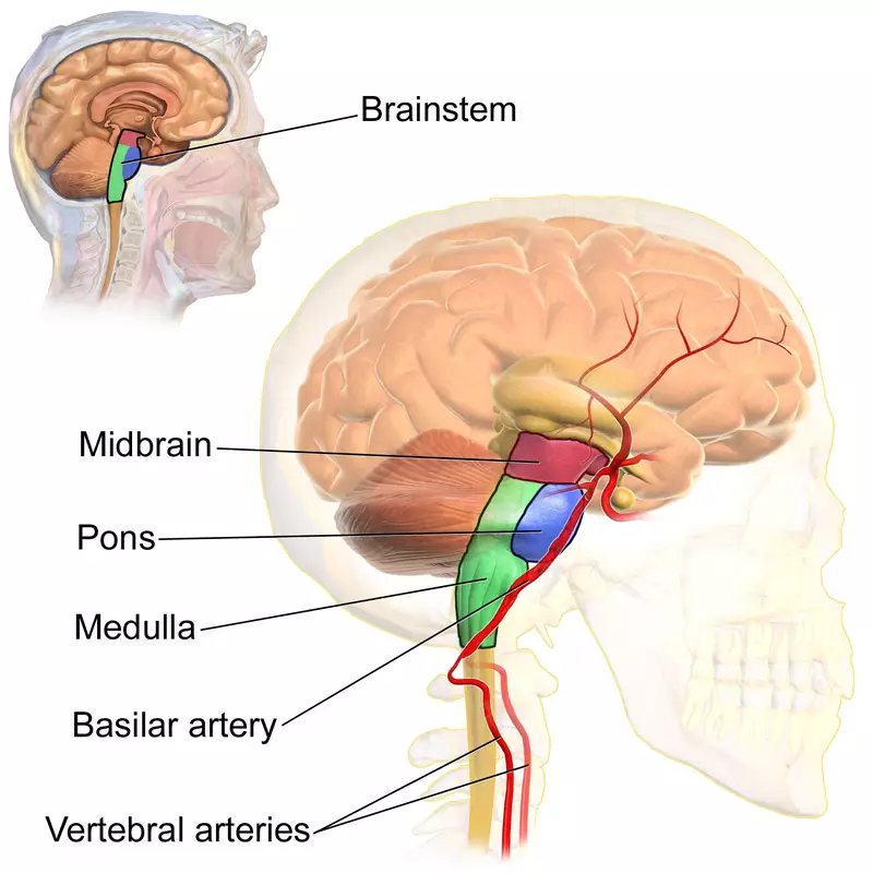
- The branches of the basilar artery are:
- Arteria spinalis anterior (ASA) anterior spinal artery
- Arteria spinalis posterior (ASP) posterior spinal artery
- Arteria cerebelli superior (SCA)
- Arteria cerebelli anterior inferior (AICA) anterior inferior cerebellar artery
- Arteria cerebelli posterior inferior (PICA) posterior inferior cerebellar artery
- Ramni meningi arteriae vertebralis
From here the basilar artery branches into the ACP (however, in 5-25% of people the ACP comes from the carotid artery).
- The ACP The posterior cerebral anteria (ACP) or posterior cerebral artery, just at the bottom of the circle of Willis, supplies blood to:
- the medial occipital lobe: occipital lobe
- the inferior and medial parts of the temporal lobe
- the limbic system
- thalamus
The primary function of the occipital lobe is vision. A stroke in the ACP commonly causes visual deficits, especially contralateral homonymous hemianopia (visual loss).
- Posterior communicans artery that connects the internal carotid artery with the artery in the circle of Willis on both the left and right
Circle of Willis
Many cerebral arteries are part of a vascular circuit of cerebral blood vessels that really run in a circle: the circle of Willis (ring of Willis). This ring or circle connects the blood vessels of the left side to the right side and the anterior to the posterior circulation. The circle of Willis is located at the base of the brain.
This blood vessel circle forms a safety mechanism because if blood flow is lost in one part, the other side can take over. However, not everyone has this security ring installed properly. About 30% of people have a complete circle of Willis.
The blood vessels that belong to the circle of Willis are:
- Arteria cerebri posterior (ACP) or posterior cerebral artery
- Arteria cerebri anterior (ACA) or anterior cerebral artery
- Arteria cerebri media (ACM) or middle cerebral artery
- Arteria communicans anterior (ACOM) or anterior communicating artery
- Arteria carotis interna (ICA) or internal carotid artery
- Arteria communicans posterior (PComm) or posterior communicating artery
More information about the circle of Willis is on this wikipedia page.
The brain seen from below
The cerebral blood vessels we are looking at in the images below lie in the confined space between the floor of the cranial cavity and the bottom of the brain.
In the image below you can see Willis' circle at the top. The names of the arteries are either on the left or on the right, but the arteries can be seen on both sides. Arteria is abbreviated to A. The top images show the same view, but with the brain drawn from below.
It looks like a "doll" pattern with "legs". The circle of Willis is actually a hexagon of blood vessels in the base of the head.
front of the forehead
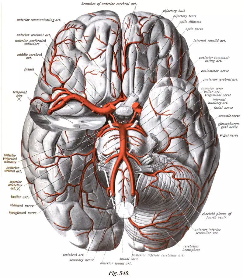
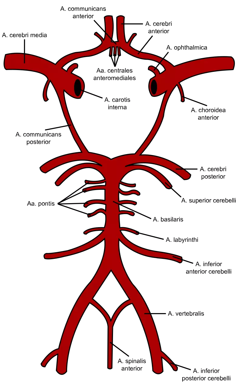
Image credits: Rhcastilhos - File:Gray519.png, Public domain, https://commons.wikimedia.org/w/index.php?curid=6187585
The brain regions that supply these blood vessels with blood:
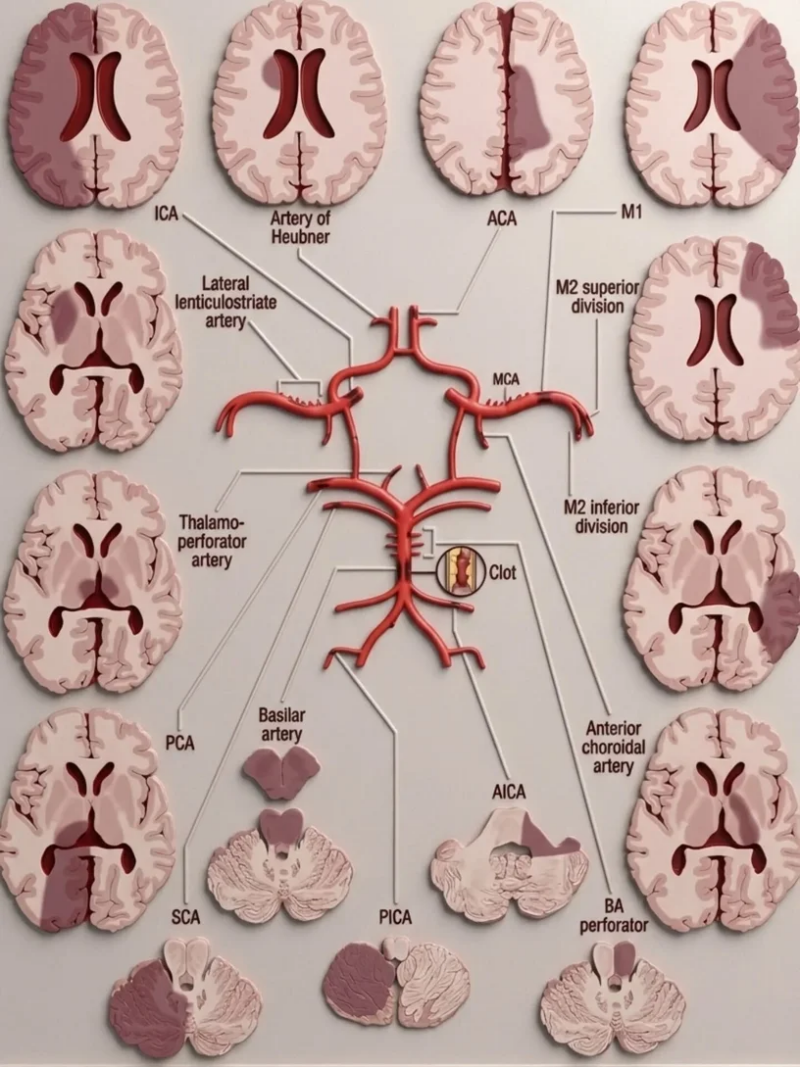
Author unknown. According to Google pictures there is no specific author of this diagram/ could not be definitively identified based on the available information. The image appears to be widely
shared, potentially originating from a source like "The Radiologist - CVS vascular territories" on Facebook.
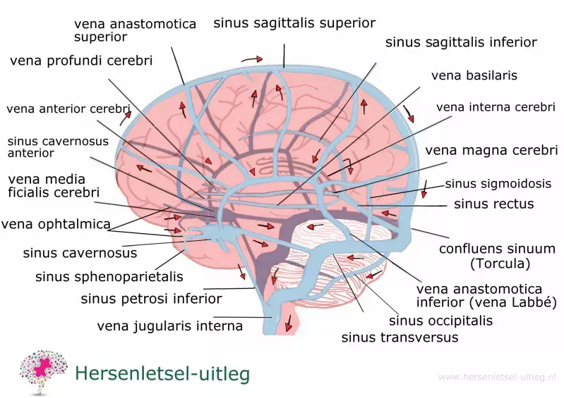
The veins of the brain, the venous drainage area
The drainage of deoxygenated blood, carbon dioxide, lactic acid and other waste products runs through veins. The veins in the brain have two systems; a deeper and a superficial system.
Deeper system
The deeper system of veins drains deoxygenated blood from the basal ganglia and structures located deep in the brain and converges behind the midbrain (mesencephalon).
Superficial
The brain veins run through cavities (sinuses) located on the surface of the brain, under the skull. The cavities are formed by the two layers of the dura mater:
- the periosteal layer, on the side of the bone. It forms a whole with the periosteum of the skull
- the meningial layer, on the side of the brain
There are blood vessels in a number of places between the two layers.
In several places the inner layer loosens and bends inward. This creates folds (dural folds) and spaces (dural venous sinuses) between the inner and outer layer.
The sinuses open into a junction of veins: confluens sinuum (from con, together; flŭĕre, to flow), where the following veins merge:
- sinus sagittalis superior
- sinus rectus
- sinus occipitalis
- left and right transverse sinus
After which the oxygen-poor blood flows further and ends in the internal jugular vein (this runs in the neck).
The superficial venous system depicted:
In case you are aware of your complaints or your failure, you may be able to find out which blood vessel is involved and vice versa.
Download the diagram here with possible complaints in case of failure of the specific cerebral artery:
Blood circulation in the skull
The bony skull doesn't receive blood from any of the four cerebral arteries, but from the middle meningeal artery.
Blood flow to the scalp
The scalp receives blood from the posterior auricular artery (near the ear), occipital artery (near the back of the head) and from the superficial temporal arteries (near the temples). In addition, the scalp receives blood from the two branches of the internal carotid artery, supra-orbital and supratrochlear.
Resources
- Adams RD, Victor M, Ropper AH. Cerebrovascular Disease. In: Adams RD, Victor M, Ropper AH, editors. Principles of Neurology. New York: McGraw-Hill, Health Professions Division, 1997: 777-873.
- Bogousslavsky J, Regli F. Anterior cerebral artery territory infarction in the Lausanne Stroke Registry. Clinical and etiologic patterns. Arch Neurol 1990; 47(2):144-150
- Bastiaanssen, C.A. & Jochems, A.A.F. (1998). Anatomie en fysiologie (4de druk). Houten: Bohn Stafleu Van Loghum
- Campbell A, Brown A, Schildroth C, et al. The relationship between neuropsychological measures and self-care skills in patients with cerebrovascular lesion. J Natl Med Assoc 1991; 83: 321324.Crossman AR, Neary D. Neuroanatomy: an illustrated colour text (2ndEd). Churchill Livingston, Harcourt Publishers Limited, London, England 2000.
- His (1895). Die anatomische Nomenclatur. Nomina Anatomica. Der von der Anatomischen Gesellschaft auf ihrer IX. Versammlung in Basel angenommenen Namen. Leipzig: Verlag von Veit & Comp.
- Kessels, R. , Eling, P. , Ponds, R., Spikman, J. & Zandvoort, M. van (Red.) (2012). Klinische neuropsychologie. Amsterdam: Uitgeverij Boom.
- Kiernan JA. Blood Supply of the Central Nervous System. In: Kiernan JA, editor. Barr's The human nervous system: an anatomical viewpoint. Philadelphia: Lippincott-Raven, 1998: 439-455
- Federative Committee on Anatomical Terminology (FCAT) (1998). Terminologia Anatomica. Stuttgart: Thieme
- Wolters, E.C. & Groenenwegen, H.J. (1996). Neurologie. Structuur, functie en dysfunctie van het zenuwstelsel. Houten/Diegem: Bohn Stafleu Van Loghum.
Recommended website: