Brodmann areas

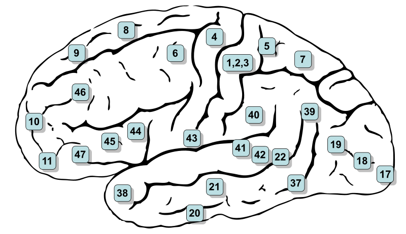
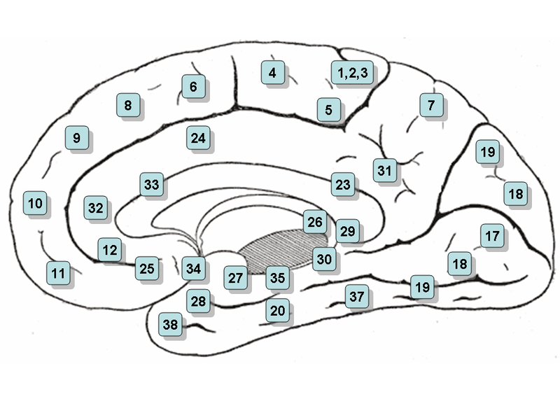

Introduction
The Brodmann brain areas, named after the German anatomist neurologist Korbinian Brodmann, give a division of the cerebral cortex into 52 numbered areas based on their structure and function.
Dr. Brodmann did a lot of research on brain cell structure.
He stained the cells under the microscope using the Nissl method for cell staining and eventually numbered areas that had similarities.
He published about his knowledge in 1909.
This classification, developed in the early 20th century, is still used in neuroscience to identify different parts of the brain.
Each area has specific functions, such as motor control, language, sensory perception or visual processing.
For example, area 4 is known for its role in controlling movement (motor control), while area 17 is essential for processing what we see (visual information).
Brodmann’s work has made a major contribution to our understanding of brain function and is often used in modern brain research techniques, such as fMRI and PET scans.
The Brodmann areas differ by:
- the construction of the brain cells in size, shape, structure, distribution and biochemical activity (cytoarchitecture)
- the microscopic anatomy of the structure and function
(histological structure) - the organization of cells
The Brodmann classification
The Brodmann areas are numbered parts of the cerebral cortex.
The classification is based on differences in the microscopic structure of brain cells. Below are the areas and their main functions.
- 1, 2, 3 - Primary somatosensory cortex (postcentral gyrus) works with information from the body
- 4 - Primary motor cortex: control of voluntary movements
- 5 – Somatosensory association cortex: combining
sensory information - 6 – Premotor cortex: preparing and planning
movements - 7 – Parietal association cortex: combining sensory information
and spatial information - 8 – Frontal visual field: control of voluntary
eye movements - 9 - Dorsolateral/anterior prefrontal cortex for motor planning and organization
- 10 – Anterior prefrontal cortex: complex decision-making, weighing goals and planning
- 11 – Orbitofrontal cortex: reward, emotions, and decision-making
- 12 – Orbitofrontal cortex: behavioral regulation and reward processing. The demarcation differs between humans and other primates.
- 13 – Insular cortex: processing of bodily signals and autonomic functions. Primarily described in primates
- 14 – Medial orbitofrontal cortex: processing of reward and bodily signals. Primarily described in primates
- 15 – Not established as a separate area in humans
- 16 – Insular cortex: processing of bodily sensations, taste, and other signals
- 17 – Primary visual cortex: initial processing of information from the eyes
- 18 – Secondary visual cortex: further processing of images
- 19 – Visual association cortex: processing of shape, movement, and spatial information, among others
- 20 – Inferior temporal gyrus: recognizing objects and visual information
- 21 – Middle temporal gyrus: language and meaning processing
- 22 – Superior temporal lobe gyrus: higher processing of sound and, especially on the left, spoken language
- 23 – Posterior gyrus: memory and processing of internally directed information
- 24 – Anterior gyrus: attention, motivation, emotions, and behavioral regulation
- 25 – Subgenual cortex: involved in mood and autonomic regulation
- 26 – Ectosplenial cortex: part of brain networks involved in memory
- 27 – Presubicular cortex: spatial orientation and memory
- 28 – Entorhinal cortex: important connection between the hippocampus and other brain regions involved in memory functions
- 29 – Retrosplenial cortex: memory and spatial orientation
-
30 – Retrosplenial cortex: memory and navigation
-
31 – Posterior engorgement: memory, attention, and self-directed information processing
-
32 – Anterior engorgement: decision-making, attention, and regulation of behavior
-
33 – Part of the anterior engorgement: processing of emotions and pain
-
34 – Dorsal entorhinal cortex: memory and olfactory information
-
35 – Perirhinal cortex: recognition of previously perceived information and memory
-
36 – Ectorhinal / perirhinal cortex: recognition of information and memory
-
37 – Occipitotemporal cortex, including the fusiform gyrus: involved in recognizing faces, among other things
-
38 – Anterior tip of the temporal lobe: meaning processing, memory, and social information
-
39 – Angular gyrus: language, reading, arithmetic, and meaning processing
-
40 – Supramarginal gyrus: language processing and
combining sensory information -
41 – Primary auditory cortex: initial processing of sound
-
42 – Auditory association cortex: further processing of sound, read also about auditory stimulus processing
-
43 – Subcentral cortex: including taste processing
-
44 – Part of Broca's area: involved in
speech production and language processing -
45 – Part of Broca's area: language production and
meaning processing -
46 – Dorsolateral prefrontal cortex: working memory, attention, and
planning -
47 – Inferior frontal cortex: meaning processing, language
and behavioral regulation -
48 – Retrosubicular cortex: historical numbering, not
generally recognized as a separate human Brodmann area -
49 – Parasubicular cortex: historical numbering, mainly
described in other mammals -
50 – No generally recognized separate human Brodmann area.
-
51 – No generally recognized separate human
Brodmann area -
52 – Parainsular cortex: transitional area between the insula and the temporal lobe
Additionally: areas 22, 39, and 40 are involved in language comprehension.
Read more about receptive aphasia and Wernicke's area.
The Brodmann areas differ in the microscopic structure of the cerebral cortex (cytoarchitecture), including the size, shape, density, distribution, and organization of nerve cells in the different layers.
Read more about the consequences per brain area via this link.
Read more about the motor cortex.
advertisements are not ours
Resources
Images by Henry Vandyke Carter - Henry Gray (1918) Anatomy of the Human Body (See "Boek" section below)Bartleby.com: Gray'sAnatomy, Plaat 726, Publiek domein, https://commons.wikimedia.org/w/index.php?curid=620390
Picture of Brodman area's by Mark Dow. Research Assistant Brain Development Lab, University of Oregon. - http://lcni.uoregon.edu/~dow/Space_software/renderings.html
Public Domain, https://commons.wikimedia.org/w/index.php?curid=8324220
Economo, C.; Koskinas, G.N. (1925). "Die Cytoarchitektonik der Hirnrinde des erwachsenen Menschen" (in German). Wien & Berlin: Springer.Peng K, Steele SC, Becerra L, Borsook D. Brodmann area 10: Collating, integrating and high level processing of nociception and pain. Prog Neurobiol. 2018Feb;161:1-22. doi: 10.1016/j.pneurobio.2017.11.004. Epub 2017 Dec 2. PMID: 29199137; PMCID: PMC5826795.Premji A, Rai N, Nelson A. Area 5 influences excitability within the primary motor cortex in humans. PLoS One. 2011;6(5):e20023. doi:10.1371/journal.pone.0020023. Epub 2011 May 16. PMID: 21603571; PMCID: PMC3095637.
Stinnett TJ, Reddy V, Zabel MK. Neuroanatomy, Broca Area. 2023 Aug 8. In: StatPearls [Internet]. Treasure Island (FL): StatPearls Publishing; 2025 Jan–.PMID: 30252352.
Tennant VR, Harrison TM, Adams JN, La Joie R, Winer JR, Jagust WJ. Fusiform Gyrus Phospho-Tau is Associated with Failure of Proper Name Retrieval inAging. Ann Neurol. 2021 Dec;90(6):988-993. doi: 10.1002/ana.26237. Epub 2021 Oct 12. PMID: 34590340; PMCID: PMC8639714.