Motor cortex
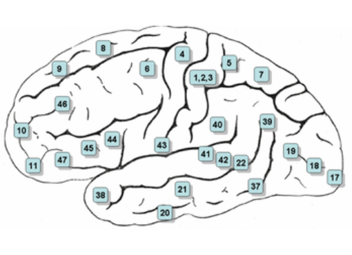
The motor cortex (movement cortex) is located in the frontal lobe and is subdivided into:
- The premotor cortex is responsible for programming movements. On the image numbered 6 (Brodmann area 6).
- The primary motor cortex is responsible for the execution of movements. On the image numbered 4 (Brodmann area 4, sometimes also called M1).
Primary, secondary, and tertiary motor cortex
The motor cortex is divided into three major areas: the primary, secondary, and tertiary motor cortex.
Each of these areas has unique functions and responsibilities.
The primary motor cortex, also known as the M1 region, is located in the frontal lobe of the brain and is directly responsible for initiating voluntary movements.
Here, signals are generated that activate the muscles to perform specific movements, such as lifting an arm or walking.
The secondary motor cortex (also known as the premotor cortex) plays an important role in planning and coordinating complex movements. This area helps integrate sensory information, enabling us to perform fine motor skills, such as writing or playing the piano.
Finally, the tertiary motor cortex is involved in the higher cognitive functions that influence movement. This area ensures that we are aware of our posture and movement in space, and enables us to adapt movements based on previous experiences and learning processes.
In short, the primary, secondary, and tertiary motor cortex work together to ensure our movements are effective and smooth.
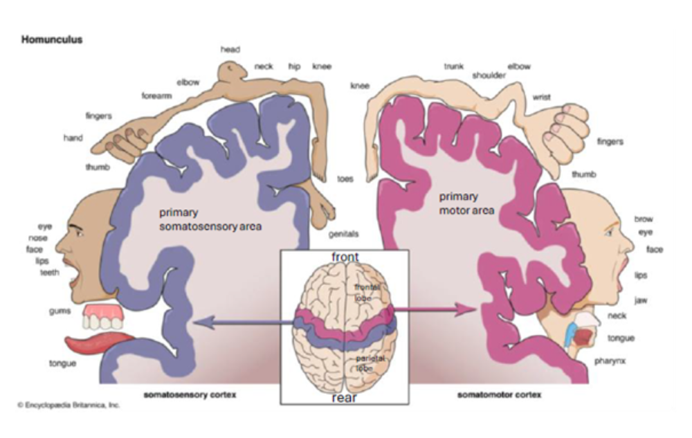
Homunculus (the motor part is pink and the sensory part is purple)
In the image below, the primary motor cortex (on the right) is shown in pink.
This representation of body parts in the motor and sensory cerebral cortex is called a homunculus. Homunculus is Latin for 'little man'. You can see a funny, little man in the image.
The motor homunculus is an overview showing which parts of the brain are responsible for the movement of certain body parts. It indicates, in proportion, how many nerve cells are involved with specific body areas.
Some body parts, such as the hands and the face, are depicted larger. This means that we have better control and precision over them. In comparison, the legs and feet are depicted smaller, which shows that we have less precise control over movements there.
This representation illustrates not only the number of muscles, but also the complexity and fineness of the movements we perform.
The benifit of the homunculus
Understanding the motor homunculus may help patients see how their movements are controlled by the brain. This also reveals which parts of the body may be vulnerable to conditions such as stroke or neurological diseases.
They can notice how treatments and exercises help their muscles and nerves.
Moreover, it makes it easier for patients to talk to their healthcare providers about their problems and expectations. Are the expectations realistic? Are the expectations too high?
This ensures a more personalized and effective treatment.
Apraxia
A particular form of apraxia, which manifests as difficulty performing everyday actions, is known as left-sided ideomotor apraxia. In ideomotor apraxia, the person understands what an object is for but cannot devise the correct movements associated with it. This may result from damage to the corpus callosum or to the right premotor area.
Injury to the prefrontal and premotor cortex and the left inferior parietal lobe may lead to ideatory apraxia. A person suffering this has difficulty remembering the correct sequence of actions.
advertisements are not ours