Cerebral hemorrhage - hemorrhagic stroke
General information about cerebral hemorrhage
Cerebral hemorrhage: When a rupture occurs in a blood vessel this causes a bleeding which presses on brain tissue. That area does not get oxygen and nutrition anymore and consequently dies. 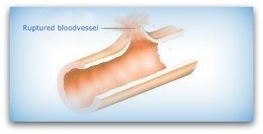
STROKE, A COLLECTIVE NAME
A stroke is a collective name for cerebral hemorrhage and cerebral infarction and stroke is also called CVA, which stands for Cerebral Vascular Accident
Loosely translated, this is an accident in the blood vessels of the brain. This can vary from a brain hemorrhage, TIA to cerebral infarction.
In stroke suddenly a portion of the brain cells do not work anymore. The person will then loss of function, such as a crippled arm, a crooked mouth or the person can not talk. A stroke can also be caused by a cerebral infarctation.
On this page we only treat the stroke by the rupture of a cerebral blood vessel; a cerebral hemorrhage.
The cause of a stroke is a tear in a blood vessel. It then flows blood in or around the brain. The blood accumulates and pushes the brain tissue away.
As a result, the brain tissue damages. A stroke can be caused by a flaw in the blood vessels (aneursyma) or malformation of the blood vessels (arteriovenous malformation AVM) or high blood pressure.
But bleeding may also be caused by an accident.
An aneurysm is a weak spot in a blood vessel that occurs throughout life. With age the aneurysm grows slowly into a bubble. High blood pressure and arteriosclerosis accelerate this process. If the aneurysm ruptures creates a hemorrhage.
WHERE IS THE BLEEDING?
Some say brain injury victims that they have suffered a stroke, another might tell you that she has had a subarachnoid hemorrhage..
What's the difference? Maybe the doctor has mentioned the term epidural hemorrhage.
To explain the difference, it is important to know how the protective layers around the brain are built.
The meninges
The brain is covered by the meninges and are the membranes that envelop the central nervous system. These brain membranes play a role in the nutrition of the brain and also serve as a pad for the protection of the brain. The meninges are built from the outside in: 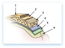
1) Skin 2) Periosteum 3) Bone 4) Dura mater hard meninges 5) Arachnoid = spider web like middle layer 6) Pia mater
Bleeding is thus divided into:
- Intracerebral hemorrhage = bleeding in the brain (in the figure above, the pink part under 6)
An intracerebral hemorrhage is bleeding in the brain caused by the rupture of a blood vessel within the brain. These injuries occur after a trauma literally tears axons in the brain's white matter. Axons are the connections that carry electrical impulses, or messages, from the neurons in the brain to the rest of the body. When this connection is sheared, serious brain damage can result because the neurons can no longer communicate. Internal bleeding can occur in any part of the brain. The bleeding may be isolated to part of one hemisphere, called a lobar intracerebral hemorrhage, or the bleeding may occur in other brain structures, such as the thalamus, basal ganglia, pons, or cerebellum.
- Subdivided in causes like:
-
- hypertension/ uncontrolled high blood pressure (BP)
- trauma
- bleeding disorder such as hemofilia
- vascular malformation such as aneurysms and Arteriovenous Malformation (AVM)
- amyloid angiopathy aqcuired dissease that weakens bloodvessel walls
- drug abuse especially cocaine or amphetamines
- SAH = subarachnoid hemorrhage, bleeding in the space between the soft meninges and arachnoid called the subarachnoid space. (in the picture above, so between layer 5 and layer 6)
-
-
aneurysm rupture ( in Circle of Willis)
-
smoking
-
alcohol
-
hypertension / high blood pressure (BP)
-
genetic risk
-
sympathomimetics drugs such as diet pills, cocain amphetamin etc.
-
estrogen deficiency (menopausal females)
-
antocoagulation
-
possibly statin medicins (anticholestoral)
-
- Subdural hemorrhage = bleeding between the dura mater and the arachnoid is bleeding as a result of an accident, usually with head injuries. (in the figure above; between layer 4 and layer 5)
- Epidural hematoma /epidural hemorrhage, bleeding outside the dura mater, between the dura and the skull. Often with a head trauma as the cause. This is often an arterial bleeding. (in the picture above intermediate layer 4 and layer 3)
When examined by a CT scan the difference is clearly.
VARIOUS CAUSES
Bleeding in the brain (intracerebral hematoma) can have various causes, such as high blood pressure (hypertension), vascular abnormalities and brain tumors.
Subdural hemorrhage (between layer 4 and 5) and epidural hemorrhage (between layer 3 and 4) are often the result of an accident (trauma).
A protrusion of an artery caused by a weak spot in the vessel wall, also known as aneurysm, often resulting in a subarachnoid hemorrhage (intermediate layer 5 and 6).
The words hematoma and hemorrhage are usually used interchangeably. Hematoma is literally bruising.
SUMMARY
If a blood vessel bursts in the brain, it bleeds in this brain area. The brain cells in that area then can't get enough oxygen or nutrients and die.
The liberated blood creates more pressure / swelling in the brain. Bleeding may be due to hypertension or congenital vascular disease, atherosclerosis, aneurysm.
On the site of the stroke center you can find more information about strokes or hemorrhages.
THE EFFECTS OF AN HEMORRHAGIC STROKE
The effects of a stroke stroke can be great:
Neurological effects as a cripple on one side of the body (hemiparesis), or an arm or leg, or even a complete paralysis (hemiplegia), but also a face half palsy (facial palsy). Often in the early stages the muscle weakness is characteristic. Later on often are seen coordinationproblems and spontaneous contraction (spasm or spasms) of the muscles which can be very annoying.
- Loss of sensory registration. The sense doing it yet, but the receipt by the brain is disrupted; sometimes reinforced, sometimes less.
- Epilepsy
- Aphasia; Usually when the hemorrhage was in the left hemisphere, because in 90% of humanity the language-area is situated there. Current research suggests that right-handers and about 70% of left-handers utilise the left brain for speech.
The aphasia (a = not, phasia = speaking) can present itself as difficulty in speaking (Broca's aphasia) as also in a disrupted language comprehension (listening and reading) Wernicke's Aphasia.
Often both forms occur at the same time = global aphasia. Aphasia is an acquired communication disorder that impairs a person's ability to process language, but does not affect intelligence. Aphasia impairs the ability to speak and understand others, and most people with aphasia experience difficulty reading and writing.
- Impairment in spatial information. Often this will happen after a hemorrhage in the right hemisphere. Getting lost in rooms, buildings, street etc. Not recognizing areas. Struggling with new info.
- Invisible effects as cognitive disorders. See also the list on the subpage "invisible effects of brain injury"