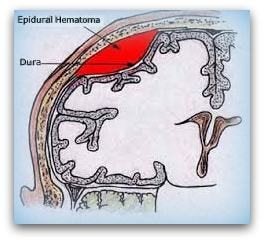
Epidural hematoma
Epidural means outside the dura, and hematoma means mass of blood. Alternative names are: Extradural hemorrhage, Extradural hematoma.
The most common cause of (intracranial) epidural hematoma is traumatic, although spontaneous hemorrhage is known to occur. Hemorrhages commonly result from acceleration-deceleration trauma and transverse forces.
Epidural hematoma may be intracranial (EDH) or spinal (SEDH). Intracranial epidural hematoma occurs in approximately 2% of patients with head injuries and 5-15% of patients with fatal head injuries. Intracranial epidural hematoma is considered to be the most serious complication of head injury.
Epidural means outside the dura, and hematoma means mass of blood.
It is a pocket of blood that forms between the skull and the tough outer layer of the brain’s protective cover, called the dura mater.
Epidural hematomas are not as common as subdural hematomas, and are most often the result of bleeding from higher-pressure arteries. The more common subdural hematomas result from bleeding of lower pressure veins.
The bleeding forms a pocket of blood that increases the pressure inside the skull. As the pocket of blood, or hematoma, grows, the pressure keeps getting higher and pushes on the brain.
This pressure can damage the brain, and in some cases even push part of the brain through the hole in the bottom of the skull that the spinal column passes through. This is called herniation, which is likely to be fatal.
Epidural hematomas may present with a lucid period immediately following the trauma and a delay before symptoms become evident. After the epidural hematoma begins collecting, it starts to compress intracranial structures.
This can be seen in the physical exam as a fixed and dilated pupil on the side of the injury. Other manifestations will include weakness of the extremities on the opposite side as the lesion (except in rare cases), and a loss of visual field opposite to the side of the lesion. The most feared event that takes place is the transtentorial, or uncal herniation which results in respiratory arrest since the medullary structures are compromised.
As with other types of intracranial hematomas, the blood may be removed surgically to remove the mass and reduce the pressure it puts on the brain.
The hematoma is evacuated through a burr hole or craniotomy.
Causes:
-
Trauma
-
Anticoagulation
-
Thrombolysis
-
Lumbar puncture
-
Epidural anesthesia
-
Coagulopathy or bleeding diathesis
-
Hepatic disease with portal hypertension
-
Cancer
-
Vascular malformation
-
Disk herniation
-
Paget disease of bone
-
Valsalva maneuver
-
Hypertension
-
Chiropractic manipulation
Click here to view the pdf