Thalamus

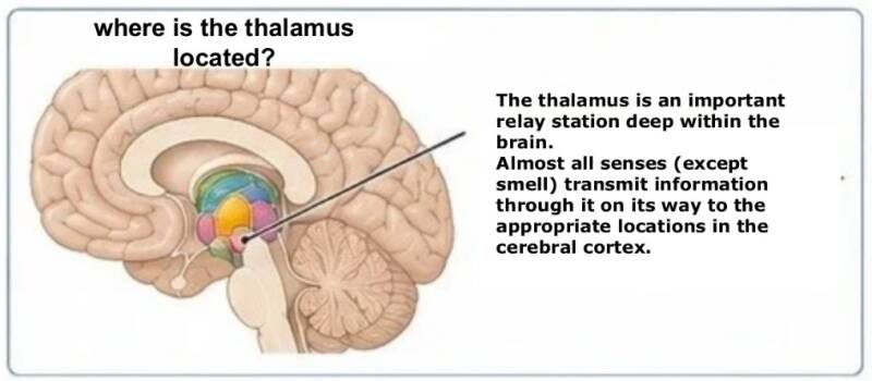
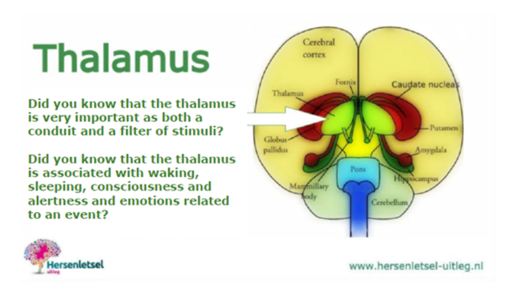
The thalamus, the yellow section on the drawing above, is an important brain nucleus and is considered one of the intermediate brain, together with the pituitary gland and the hypothalamus. It is the most 'wired' portion of the brain.
Introduction
Foreword: The brain works together as a single unit. Brain functions are distributed throughout the brain regions and arise from the exchange between the regions. Nevertheless, symptoms can be identified per brain region.
Do you only want the short summary? Then follow this link.
Do you only want to read the possible symptoms per core group and per adjacent brain regions? Follow this link.
Do you want to know more about the anatomy, functions, and potential damage per core group? Then follow this link.
Thalamus: most wired part of the brain
The thalamus is an important relay station deep within the brain. Virtually all sensory information – such as what you see, feel, and hear – is transmitted via the thalamus to the cerebral cortex.
It helps to focus attention on important information and filter out less important stimuli.
It also assists with movement, memory, emotions, and with being conscious, awake, and alert.
It plays a role in akinetic mutism. This is a syndrome, a consequence of brain injury, in which the person speaks little or not at all (mutism) and moves little or not at all (akinesia). The person may follow you with their eyes but otherwise appears apathetic and does almost nothing of their own accord.
Thalamus is derived from the Greek word for hidden central inner chamber or bedroom; θάλαμος, because it lies deep within the brain and forms the 'central' chamber.
It is a deep-seated central nucleus in the brain
- virtually all sensory information (except smell) enters it first
- it distributes and organizes information before it goes to the cerebral cortex
- it is in contact with almost all parts of the cortex via thousands of connections
You can therefore view the thalamus as the central inner chamber where information first enters and is forwarded
Senses
l
Thalamus
(central inner chamber)
l
Cerebral Cortex
Gateway to the Cerebral Cortex
The connections from the thalamus to the cerebral cortex return from the cortex to the thalamus.
This is called the thalamo-cortico-thalamic circuit. As a result, minor thalamic lesions can mimic cerebral cortex syndromes, and in some syndromes, the specialization of the cerebral hemisphere can be recognized.
As a gateway to the cerebral cortex, the thalamus has to transmit many sensory stimuli (except smell), but it must also filter stimuli, allowing healthy individuals to "shut off" certain stimuli. It is therefore one of the so-called "filters" (stimulus filters), a control center to help the brain focus on important and unimportant stimuli.
The main core groups of the thalamus and adjacent structures (in the diencephalon)
Important to know:
The thalamus works closely with other brain regions. The symptoms a person experiences may correspond to damage to a specific core group, but are often also determined by the cooperation between different brain networks.
The following anatomical terms explain the names of the core groups:
The image below is a schematic anatomical representation. The exact shape, size, and mutual boundaries of the core groups may vary slightly depending on the anatomical cross-section and the anatomical reference work.
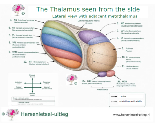
Discussion of the image of the thalamus viewed from the side (lateral view) with the adjacent metathalamus
The image shows the main nuclear groups of the thalamus in a lateral view. The thalamus consists of various nuclear groups that receive, process, and transmit information to the cerebral cortex. Not all nuclear groups are fully visible from this view. Therefore, hidden or partially hidden structures are indicated with a curved gray arrow, while straight black arrows point to structures that are directly visible.
The lamina medullaris interna (Y-shaped white matter tract) divides the thalamus into different anatomical nuclear groups.
The intralaminar nuclei are located within this white matter tract. The midline nuclei lie directly along the third ventricle and are barely visible from a lateral view. The reticular nucleus forms a thin layer on the outside of the thalamus and plays an important role in regulating the information processed by the thalamus.
Located at the posterior side are the lateral knee-form body (LGN (Medial Geniculate Nucleus)) and the medial knee-form body (MGN (Medial Geniculate Nucleus)). Together, they form the metathalamus. The LGN processes visual information, while the MGN is involved in processing auditory information.
The illustration is based on the anatomical classification as described in Gray's Anatomy, supplemented and verified with Mai & Paxinos and Blumenfeld.
For each section, we elaborate on the following:
- Where are the nuclei located?
- From where do they receive information?
- Where do they send information?
- What do these nuclei do?
- Possible consequences of damage
- What do you notice about this in daily life?
- Important to know
- In-depth information for healthcare professionals
- Anatomical references
The list is written in plain language (B1 level), but is based on authoritative anatomical and neurological literature, including Gray's Anatomy, Mai & Paxinos, and Blumenfeld. Where scientific insights are still developing, this is indicated in the text. In this way, we combine understandable explanations with current medical knowledge.
Two nuclei: the pulvinar at the back of the thalamus and a nucleus in the middle at the bottom, the nucleus reticularis thalami, have partially overlapping functions:
| FUNCTION | PULVINAR | NUCLEUS RETICULARIS THALAMI |
|---|---|---|
| Attention regulation | mostly visual | general and modeling |
| Visual processing | strongly involved | supports visual attention |
| Integration of sensory information | multisensory | filters unwanted information |
| Role in consciousness | less direct, more in perception | crucial for alertness and focus |

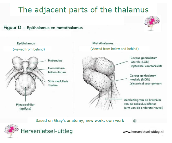
Anatomical note
The metathalamus and the epithalamus do not belong to the dorsal thalamus. They are included in this list nonetheless, because according to the anatomical atlases of Gray's Anatomy, Mai & Paxinos, and current clinical neurological knowledge of Blumenfeld, they are described together with the thalamus as parts of the diencephalon (interbrain).
- Gray's Anatomy: The Anatomical Basis of Clinical Practice
(most recent edition) - Mai JK & Paxinos G. Atlas of the Human Brain
- Blumenfeld H. Neuroanatomy Through Clinical Cases
Brief summary
Why is the thalamus important?
If a core group does not function properly, it may lead to:
- problems with movement
- altered or diminished sensations
- problems seeing or ignoring things
- difficulty with thinking, planning, and memory
- pain complaints that cannot be easily explained (such as thalamic pain)
Important to know
The thalamus contains more than fifty small nuclei.
The classification into fifteen nuclear groups helps to better understand the functions associated with complaints and to understand the structure and function of the thalamus.

The thalamus houses more than 50 distinct nuclei, which are organized into functionally diverse core groups. Each group is linked to specific symptoms that can occur upon injury. In the text above, we discuss in detail the consequences of injury to the most important core groups.
These nuclei function as relay stations, where information from other parts of the nervous system is transmitted to specific areas in the cerebral cortex. Important to know: The thalamus works closely with other brain regions. The symptoms mentioned on this page may correspond to damage to a specific core group, but are often also determined by the cooperation between different brain networks. In the text below, we discuss the symptoms per core group.
Anterior nuclei (front)
Help store new memories and learn things
Mediodorsal nucleus (MD)
Important for planning, making decisions, and executing plans
Lateral nucleus (LD)
Processes visual information and helps to recognize and see things
Lateral posterior nucleus (LP)
Processes visual information and helps to recognize and see things
Pulvinar (posterior center)
Important for visual attention, seeing movement, and linking information between different brain regions
Ventral anterior nucleus (VA)
Helps control movements via connections with the basal ganglia
Ventral lateral nucleus (VL)
Important for initiating and controlling voluntary movements
Ventral posterolateral nucleus (VPL)
Brings sensation from the body (such as touch, pressure, and posture) to the cerebral cortex
Ventral posteromedial nucleus (VPM)
Brings sensation from the face and taste to the cerebral cortex
Intralaminar nuclei (IL)
Give an ‘alarm’ and keep you awake and alert
Midline nuclei
Play a role in emotions, pain perception, and the autonomic nervous system (unconscious bodily functions)
Habenular nuclei (habenula)
Help with reward, motivation, and the dopamine regulatory system
Epithalamic nuclei (epithalamus)
Support the biological clock and the day-night rhythm
Geniculate bodies
Important for hearing (medial) and vision (dorsal)
Reticular nucleus
Thin layer surrounding the thalamus that regulates the flow of information and influences the rhythm of brain activity
The possible consequences of thalamic damage and by core group
Important to know
The thalamus works closely with other brain regions. The symptoms mentioned may be consistent with damage to a specific core group, but are often partly determined by the cooperation between different brain networks. Not everyone with thalamic damage experiences the same symptoms. Which symptoms arise depends, among other things, on:
• which core group is affected
• the extent of the injury
• whether one or more core groups are damaged
• which other brain regions are involved.
Many symptoms can also have another cause.
This list shows which symptoms can occur with damage to the thalamus or adjacent structures.
We elaborate on the following consequences:
• Memory
• Executive functions
• Attention and concentration
• Movement
• Changes in sensation and pain
• Vision
• Visual attention
• Hearing
• Sensory overstimulation
• Emotions
• Behavior
• Fatigue
• Reduced mental resilience
• Sleep
• Consciousness
• Language
• Spatial orientation
For each consequence, we refer to the explanation regarding the core groups of the thalamus as shown in the green drop-down menu above and the numbers in the image below:
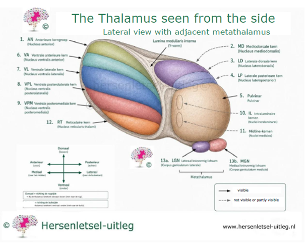
Below you can find a summary table with the main function and symptoms
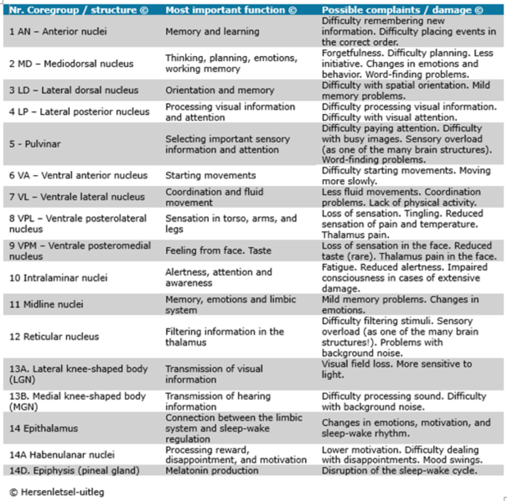
Possible consequences
Memory
Possible complaints:
- Difficulty remembering new information
→ 1. AN - anterior core group, 2. MD - Mediodorsal nucleus
- Forgetfulness
- Difficulty placing events in the correct order
- Difficulty storing experiences properly
- Mild memory problems
Attention and concentration
Possible complaints:
- Easily distracted
- Difficulty maintaining attention
- Difficulty multitasking
- Slower information processing
Movement
Possible complaints:
- Slower movement
→ 6.VA - ventral anterior nucleus
→ 7.VL - ventral lateral nucleus
- Less fluid movements
→ 7.VL - ventral lateral nucleus
- Coordination problems
→ 7.VL - ventral lateral nucleus
- Difficulty initiating a movement
→ 6.VA - ventral anterior nucleus
- Less precise movements
→ 7.VL - ventral lateral nucleus
Changes in sensation and pain
- Numbness or tingling on one side of the body
→ 8.VPL - Ventral posterolateral nucleus
→ 9.VPM - Ventral posteromedial nucleus
- Reduced sense of touch
→ 8.VPL - Ventral posterolateral nucleus
→ 9.VPM - Ventral posteromedial nucleus
- Impaired sense of temperature
→ 8.VPL - Ventral posterolateral nucleus
→ 9.VPM - Ventral posteromedial nucleus
- Impaired sense of pain
→ 8.VPL - Ventral posterolateral nucleus
→ 9.VPM - Ventral posteromedial nucleus
- Burning or severe pain without a clear cause
→ 8.VPL - Ventral posterolateral nucleus
→ 9.VPM - Ventral posteromedial nucleus
Déjerine-Roussy syndrome / Thalamic pain syndrome
Thalamic pain occurs in the thalamus following a stroke (CVA).
The pain is then felt on the opposite side of the body from where the stroke occurred, i.e., in one half of the body.
The pain can be localized or felt over a larger area in the legs, arms, or face.
It involves burning, stabbing, pricking, and tingling sensations. Changes in the weather or certain movements or activities may worsen it.
Characteristics:
- burning pain
- stabbing or shooting pain
- prickling or tingling sensations
- pain in the arm, leg, and/or face on the opposite side of the body
- pain upon light touch (allodynia)
- excessive pain response (hyperalgesia)
- aggravation by cold, heat, weather changes, movement, or stress
Read more on our page on pain.
Vision
Possible complaints:
- Difficulty processing visual information
→ 4. LP – Posterior lateral nucleus
- Difficulty surveying a busy environment
- Difficulty recognizing important visual information among many other stimuli
- Visual field defect on the opposite side of the visual field
→ 13a. LGN – Metathalamus (Lateral knee-shaped body)
Important to know
The thalamus does not enable us to see.
That primarily happens in the cerebral cortex at the back of the brain. However, the thalamus does help transmit and select visual information.
Visual attention
Possible complaints:
- Difficulty focusing attention on an object or person
- Difficulty maintaining attention while looking
→ 5. Pulvinar
→ 2. MD – Mediodorsal nucleus - Difficulty finding an object in a busy environment
- Becoming distracted more easily by movement in the environment
→ 5. Pulvinar
→ 4. LP – Posterior lateral nucleus
Important to know
Visual attention is different from being able to see well. The eyes may function normally, yet it is still difficult to focus attention on the correct visual information. This is not about sensory overload.
Hearing
Possible complaints:
- Difficulty processing sound
→ 13b. MGN – Medial knee-shaped body (metathalamus)
- Difficulty recognizing differences between sounds
→ 13b. MGN – Medial knee-shaped body (metathalamus)
- Difficulty understanding speech clearly in a noisy environment
→ 13b. MGN – Medial knee-shaped body (metathalamus)
→ 12. Reticular nucleus
→ 5. Pulvinar
Important to know
Hearing itself may be normal, while the processing of sounds in the brain is still difficult.
Sensory Overstimulation
Important to know
Sensory overstimulation concerns the burden of sensory information ánd the reduced capacity to handle sensory stimuli as a result of brain injury. Consequently, ordinary stimuli, which previously posed no problem, may be perceived as too much or too intense.
The thalamus plays an important role in this, but works closely with other brain regions. Sensory overstimulation is usually not caused by a single brain region alone. On this page, we discuss ónly the role of the thalamus, the metathalamus, and the epithalamus.
! Other brain regions that may also play a role are not discussed in further detail on this page!
Possible effects:
- Difficulty understanding speech clearly in a noisy environment.
→ 13b. MGN – Medial knee-shaped body (metathalamus)
→ 12. Reticular nucleus
→ 5. Pulvinar
- Sounds are perceived as loud or intense.
→ 13b. MGN – Medial knee-shaped body (metathalamus)
→ 12. Reticular nucleus - Difficulty with background noise
→ 12. Reticular nucleus
→ 13b. MGN – Medial knee-shaped body (metathalamus)
→ 5. Pulvinar - Light is easily perceived as bothersome.
→ 13a. LGN – Lateral knee-shaped body (metathalamus)
→ 5. Pulvinar - Difficulty with busy images or a lot of movement around you
→ 5. Pulvinar
→ 4. LP – Lateral posterior nucleus - Touch may be perceived as overwhelming more quickly.
→ 8. VPL – Ventral posterolateral nucleus
→ 9. VPM – Ventral posteromedial nucleus - Pain stimuli may be experienced more intensely.
→ 8. VPL – Ventral posterolateral nucleus
→ 9. VPM – Ventral posteromedial nucleus
Emotions
Possible symptoms:
- Less control over emotions
→ 2. MD - Mediodorsal nucleus
→ 14A. Habenular nuclei
- Reacting emotionally quickly
- Showing less emotion than before.
- Mood swings.
→ 2. MD – Mediodorsal nucleus
→ 14A. Habenular nuclei
- Reduced motivation
- Difficulty coping with disappointments
- Experiencing less pleasure in activities that used to be fun
Important to know
Emotions arise from the cooperation of different brain regions. The mediodorsal nucleus and the habenular nuclei play an important role in this, but work closely with, among others, the prefrontal cortex and other parts of the limbic system.
Behaviour
Possible complaints:
- Taking less initiative
- Difficulty with planning
→ 2. MD – Mediodorsal nucleus - Difficulty starting an activity independently
→ 2. MD – Mediodorsal nucleus
→ 6. VA – Ventral anterior nucleus (when initiating movements is involved) - Changes in social behavior
→ 2. MD – Mediodorsal nucleus - Less ability to respond flexibly to changes
→ 2. MD – Mediodorsal nucleus
Important to know
Changes in behavior do not mean that a person's personality changes completely. Often, it involves subtle changes in initiative, planning, social interaction, and adapting to new situations.
Sleep
Possible symptoms:
- Disruption of the sleep-wake rhythm
→ 14. Epithalamus
→ 14B. Pineal gland - Difficulty falling asleep or staying asleep
→ 14B. Pineal gland (when melatonin balance is disrupted) - Becoming tired more quickly during the day
→ 14. Epithalamus
→ 10. Intralaminar nuclei (when alertness is also reduced)
Important to know
The sleep-wake rhythm is regulated by multiple brain regions. The pineal gland produces the hormone melatonin and is influenced by signals from various brain networks. The epithalamus plays a supporting role in this.
Consciousness and alertness
Possible symptoms:
- Becoming drowsy more quickly
→ 10. Intralaminar nuclei - Being less alert
→ 10. Intralaminar nuclei - Difficulty staying awake and alert for extended periods
→ 10. Intralaminar nuclei - Severe disturbances of consciousness with extensive damage
→ 10. Intralaminar nuclei
Important to know
The intralaminar nuclei are part of a larger network involved in alertness and consciousness. Severe disturbances of consciousness usually only occur when multiple components of this network are damaged.
Fatigue
Possible symptoms:
- Mental fatigue
→ 2. MD – Mediodorsal nucleus
→ 10. Intralaminar nuclei - Becoming exhausted more quickly after a conversation, activity, or busy environment
→ 2. MD – Mediodorsal nucleus
→ 10. Intralaminar nuclei - Being able to sustain activities for a shorter period of time
→ 2. MD – Mediodorsal nucleus
→ 10. Intralaminar nuclei - Needing more time to recover after physical or mental exertion
→ 2. MD – Mediodorsal nucleus
→ 10. Intralaminar nuclei - Needing rest moments sooner during the day
→ 10. Intralaminar nuclei - Feeling that the brain is "full" after a lot of information
→ 2. MD – Mediodorsal nucleus
→ 10. Intralaminar nuclei
Important to know
Fatigue after a brain injury is more than just ordinary tiredness. Many people find that their brains become exhausted more quickly and require more recovery time.
The thalamus plays an important role in alertness, attention, and information processing. However, mental fatigue is usually not caused by a single brain area alone. Various brain networks work together in this process.
In this list, we discuss only the role of the thalamus. Other brain regions that also contribute to mental fatigue are not further elaborated on this page.
Reduced Mental Capacity
What does mental capacity mean?
Mental capacity is the amount of effort the brain can handle before symptoms appear. After a brain injury, this capacity can decrease. As a result, activities that previously required little effort may now demand a lot of energy.
Mental capacity consists of various components.
Two important forms are cognitive capacity and sensory capacity:
1) Cognitive capacity
Possible effects:
- Difficulty thinking for extended periods
- Shorter ability to concentrate
- Difficulty processing a lot of information at once
- Making mistakes more quickly when a task takes longer
- Difficulty performing multiple tasks simultaneously
- Needing more breaks while thinking or working
Potentially involved core groups
→ 2. MD – Mediodorsal nucleus
→ 10. Intralaminar nuclei
→ 5. Pulvinar
2) Sensory Load
Possible effects:
- Sound is quickly perceived as overwhelming.
- Background noise consumes a lot of energy.
- Light quickly becomes bothersome.
- Busy images require a lot of effort.
- Touch can quickly feel too intense.
- It is difficult to sustain multiple stimuli at once.
A long recovery time is needed after a busy environment.
Possibly involved nucleus groups
→ 12. Reticular nucleus
→ 5. Pulvinar
→ 10. Intralaminar nuclei
→ 13a. LGN – Lateral knee-shaped body (metathalamus)
→ 13b. MGN – Medial knee-shaped body (metathalamus)
→ 8. VPL – Ventral posterolateral nucleus
→ 9. VPM – Ventral posteromedial nucleus
Important to know
Cognitive and sensory capacity often influence each other. Someone who has to process many sensory stimuli may subsequently be less able to concentrate or think for a shorter period. Conversely, prolonged mental exertion can cause someone to become more sensitive to sound, light, or other sensory stimuli.
Although these forms of capacity are related, they are not the same functions. It is therefore important to distinguish them from one another.
Other problems mentioned following injury to the thalamus
Reduced taste
→ 9. VPM – Ventral posteromedial nucleus
This problem is rare.
Language problems
Possible symptoms:
- Difficulty finding a word (word-finding problems)
→ 2. MD – Mediodorsal nucleus
→ 4. LP – Posterior lateral nucleus
→ 5. Pulvinar
(occurs especially with damage to the left thalamus)
- Speaking more slowly because the correct word is not found immediately
→ 2. MD – Mediodorsal nucleus
→ 5. Pulvinar
(especially left)
- Using a wrong word when you actually mean something else
→ 2. MD – Mediodorsal nucleus
→ 5. Pulvinar
(less common)
- Difficulty constructing a sentence properly
→ 2. MD – Mediodorsal nucleus
(less common)
- Difficulty understanding spoken language well
→ 5. Pulvinar
→ 4. LP – Posterior lateral nucleus
(rare, usually with more extensive or left-sided damage)
- Difficulty following a conversation when there is a lot of background noise
→ 13B. MGN – Medial knee-shaped body (metathalamus)
→ 12. Reticular nucleus
→ 5. Pulvinar
(this is usually a problem with information processing and not with language comprehension itself)
Word finding (anomia) is by far the most common language disorder following thalamic injury.
Language production (sentence formation and fluent speech) is affected much less frequently and usually only in cases of more extensive or left-sided lesions.
Language comprehension is even rarer as a direct result of an isolated thalamic lesion.
What do you notice about this in daily life?
For example, you may notice that:
- a word is "on the tip of your tongue" but you can't get it out
- you need more time to say something
- conversations take more energy
- you sometimes use a different word than you mean
- you have trouble following a conversation when multiple people are talking at the same time
- you lose the thread of a conversation more quickly
Important to know
The thalamus is not a language center. The main language areas are located in the cerebral cortex, primarily in the left hemisphere.
However, the thalamus does play an important role in the networks that support language.
As a result, problems may arise with finding words, speaking fluently, processing language.
These symptoms are often subtle and vary from person to person.
Executive functions (goal-directed action)
Possible complaints
- Difficulty making a plan
- Difficulty performing a task step by step.
- Difficulty maintaining an overview
- Difficulty setting priorities
- Difficulty starting an activity
→ 2. MD – Mediodorsal nucleus
→ 6. VA – Ventral anterior nucleus (when initiating movements is also affected)
- Difficulty completing an activity
- Difficulty changing approach when something doesn't work
- Less ability to solve problems
- Reacting impulsively more quickly
- Less ability to assess the potential consequences of a choice
What do you notice about this in your daily life?
See our special page about this issue.
For example, you may notice that:
- it is harder to plan a day
- cooking according to a recipe takes more effort
- grocery shopping without a list becomes more difficult
- you get stuck more easily when something unexpected happens
- you need help more often to keep track
- you start a task but do not finish it
- it is harder to make choices
Important to know
Executive functions help you act in a goal-oriented manner. They enable you to plan, organize, make choices, solve problems, and adapt your behavior when a situation changes.
The mediodorsal nucleus plays an important role in this process because it works closely with the prefrontal cortex. Damage to this nucleus can make it more difficult to apply knowledge and skills in daily life, even if intelligence itself remains intact.
See our dedicated page on this issue.
Spatial orientation
For example:
- difficulty finding one's way
- difficulty maintaining an overview
- difficulty with spatial attention
especially:
Here you can download a document by Emmanuel Carrera M.D. and Julien Bogousslavsky, M.D., containing more information about thalamus injury and behavior.
advertisements are not ours
Resources
Cairns, H., RC Oldfield, JB Pennybacker, D. Whitteridge: akinetische mutisme met epidermoïd cyste op de 3e ventrikel . In: Brain . Volume 64, 1941, p 273-290.
Carrera, E., & Bogousslavsky, J. (2006). The thalamus and behavior: Effects of anatomically distinct strokes. Neurology, 66(12), 1817–1823. https://doi.org/10.1212/01.wnl.0000219679.95223.4c
Eyskens, E., Feenstra, L., Meinders, A., Vandenbroucke, J. P., & Van Weel, C. (1997). Codex Medicus (10e ed.). Maarssen, Nederland: Elsevier Gezondheidszorg.
File:Thalamus3.PNG - Wikimedia Commons. (2007, 20 januari). Consulted from https://commons.wikimedia.org/wiki/File:Thalamus3.PNG

Haaxma, R. (2013). Neurologie van cognitie en gedrag in hoofdlijnen. Houten, Nederland: Springer Media.
Herrero, M., Barcia, C., & Navarro, J. (2002). Functional anatomy of thalamus and basal ganglia. Child's Nervous System, 18(8), 386–404. https://doi.org/10.1007/s00381-002-0604-1
Hersenletsel uitleg (page on the thalamus)
Kuks, J. B. M., Snoek, J. W., Oosterhuis, H. G. J. H., & Fock, J. M. (2003). Klinische neurologie (15e ed.). Houten, Nederland: Bohn Stafleu van Loghum.
Palm, J. (2012). Omgaan met Hersenletsel. Assen, Nederland: Van Gorcum.
Schuurman, P. R., Bosch, D. A., Bossuyt, P. M., Bonsel, G. J., Van Someren, E. J., De Bie, R. M., . . . Speelman, J. D. (2000). A Comparison of Continuous Thalamic Stimulation and Thalamotomy for Suppression of Severe Tremor. New England Journal of Medicine, 342(7), 461–468. https://doi.org/10.1056/nejm200002173420703
Sherman, S. M. (2005). Thalamic relays and cortical functioning. Progress in Brain Research, , 107–126.https://doi.org/10.1016/s0079-6123(05)49009-3
Thalamus, consulted from: http://medicaltextbooksrevealed.s3.amazonaws.com/files/11185-53.pdf
Thalamus, consulted from: http://zlab.rutgers.edu/modules/teaching/docs/Thalamus/Thalamus%20Lecture.pdf
Thalamus, consulted from: http://www.neuroanatomy.wisc.edu/coursebook/thalamus.pdf
Wikipedia: https://nl.wikipedia.org/wiki/Bestand:Thalamus3.PNG

Zesiewicz, T. A., Elble, R., Louis, E. D., Hauser, R. A., Sullivan, K. L., Dewey, R. B., . . . Weiner, W. J. (2005). Practice Parameter: Therapies for essential tremor: Report of the Quality Standards Subcommittee of the American Academy of Neurology. Neurology, 64(12), 2008–2020. https://doi.org/10.1212/01.wnl.0000163769.28552.cd
Anatomic resources:
- Gray's Anatomy: The Anatomical Basis of Clinical Practice (meest recente editie)
- Mai & Paxinos – Atlas of the Human Brain
- Blumenfeld – Neuroanatomy Through Clinical Cases
"Thalamus3" by Original uploader was Albert Kok at nl.wikipedia - Originally from nl.wikipedia; description page is/was here.. Licence Public domain via Wikimedia Commons.

